기업맞춤형 기술지원, 정책뉴스, 지원사업소개, 유망기술 제공 등
다양한 기업지원 정보제공을 위해 노력하겠습니다.


| 보유기관 | 한국핵융합에너지연구원 |
|
|---|---|---|
| 보유지역 | 군산 | |
| 보유실험실 | 국가핵융합연구소 플라즈마기술연구센터 1층 103 | |
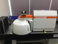
| 모델명 | inVia Raman microscope | |
| 장비용도 | ○ UV Micro-Raman Spectroscopy의 경우 Graphene에서부터 nanodiamond/MoS2 등 다양한 물질 분석 및 박막/표면 개질에 응용되고 있음 ○ 본 장비는 ‘플라즈마 융합 원천 연구사업’에서 진행되고 있는 ‘플라즈마를 이용한 나노분말의 합성, 표면개질’ 등에서 기초물성 테스트에 응용되어질 수 있음 ○ 바이오/플렉서블 디스플레이 등에 물질의 특성 분석을 위한 주요 장비로 응용 될어질 수 있음. ○ 현재 본 연구소에서 수행중인 양자점 태양전지 개발에 있어 양자점의 특성 및 효율 분석에 직접적으로 응용되어짐. ○ 농식품 포장재 특성연구에 활용 |
|
| 주요사양 | ○ UV Micro-Raman spectroscopy는 다음과 같은 장점을 보유하고 있다. 1. Sensitivity 라만 분광 (Raman Scattering) 신호는 짧은 파장의 레이저를 투과할 때 증가한다. 2. Avoiding fluorescence 라만 분석 시 나타나는 형광 (Fluorescence) 현상은 가시광선 (visible region) 영역의 전자기 스펙트럼 (electromagnetic spectrum)에서 주로 일어나기 때문에 짧은 파장의 라만 분광은 나노물질 및 박막 분석시 필수적이다. 3. Resonance Raman 라만 레이저의 파장과 시료의 흡수 파장이 동시에 일어날 경우 공명 (resonance)가 일어난다. 따라서 다양한 파장의 라만 분광을 통해 선택적 미량분석에 응용된다 플라즈마 융합 원천 연구사업에는 플라즈마를 활용한 나노물질의 합성, 스퍼터링 (sputtering)을 통한 박막 형성, 그리고 플라즈마를 통한 표면 개질에 관한 연구를 수행 주에 있다. 짧은 파장(320 nm) 라만의 경우 형광(Fluoscence) 현상이 강한 나노 다이아몬드(Nanodiamond), 그래핀(Graphene/Graphene oxide), 양자점(Quantum dot) 등 플라즈마를 이용한 물질 및 표면 연구에 필수적이며 532 nm의 파장은 320 nm 파장을 이용한 특성 연구에 그 기준점 (reference)를 제공 해줄 것이다. Renishaw inVia Spectrometer System for Raman spectral analysis using visible excitations at 325nm. Including: (USD $288,884.00) 1.1 Spectrometer Stigmatic single pass spectrograph with the following specification: 1.1.1 Extremely high efficiency 250 mm focal length spectrograph (>30% throughput in spectrograph). 1.1.2 Laser spot size fixed to 2 um (objective and excitation wavelength dependent) 1.1.3 Automated, kinematically mounted, magnetically attached, Rayleigh line rejection filter set for 325 nm excitation, allowing measurement of Raman spectrum from 250 to 4000 cm-1 from the laser line. 1.1.4 Fully optimised laser beam path optics for UV laser delivery with fixed laser spot size. 1.1.5 Fully optimised mirror, prism and optical path for UV signals. Upgrade to UV ND filters 1.1.6 Optimised UV lens set; kinematically mounted for optimised throughput and spectral resolution. 1.1.7 Most advanced continually adjustable true confocal facility using manual variable slit. 1.1.8 UV optimised 2400 or 3600 g/mm grating on interchangeable magnetic kinematic mount for 325 nm excitation. 1.1.9 Unique fast ‘extended scanning’ facility for measurement of high resolution spectra with wider wavelength range than can be accommodated on a single CCD exposure, without any ‘stitching’ of spectra together. Spectral resolution continuously variable via CCD binning control. 1.1.10 UV and NIR enhanced deep depletion CCD array detector (1024 x 256 pixels). Peltier cooled to ?70 ºC. No water or liquid nitrogen required. 1.1.11 Motorized neutral density filters offering 16 different power pixel levels from 0.00005 to 100%. 1.2 Kinematic system baseplate 1.2.1 Kinematic honeycomb baseplate for spectrometer, microscope and up to three lasers. (note: instead of this baseplate, an optical table is required, not include, for an UV system with244nm laser) 1.3 Microscope Specially adapted Research Grade Leica microscope allowing advanced true confocal measurements. Including: |
|
| 장비사용 지원신청 |
|
|


